

In this article
Melanotan I is a synthetic analogue of alpha-melanocyte-stimulating hormone, commonly abbreviated as α-MSH. It was developed to improve the stability and biological activity of the natural melanocortin peptide while preserving its ability to activate melanocortin receptors.
Melanotan I is also known scientifically as afamelanotide or [Nle4, D-Phe7]-α-MSH.
The peptide contains 13 amino-acid residues, like natural α-MSH, but two residues have been modified:
- Methionine at position 4 is replaced by norleucine
- L-phenylalanine at position 7 is replaced by D-phenylalanine
These modifications produce a peptide that is more resistant to degradation and more potent than native α-MSH in several experimental systems.
Melanotan I research commonly involves:
- MC1R receptor biology
- Melanocortin signaling
- cAMP production
- Melanocyte function
- Eumelanin synthesis
- Peptide stability
- Structure-activity relationships
- Receptor-binding studies
- Photobiology
- Analytical peptide characterization
Although afamelanotide is also the active ingredient in an FDA-approved pharmaceutical product for a specific medical indication, research-grade Melanotan I should not be treated as equivalent to an approved drug formulation.
For research purposes only. Not for human or veterinary use.
What Is Melanotan I?
Melanotan I is a synthetic peptide derived from α-MSH.
Natural α-MSH is a 13-amino-acid melanocortin peptide involved in pigmentation and several other physiological processes.
Melanotan I retains the overall α-MSH backbone but includes two important substitutions.
It is commonly represented as:
[Nle4, D-Phe7]-α-MSH
These substitutions were designed to increase peptide stability and receptor activity.
The resulting molecule is also known as NDP-MSH.
NDP-MSH stands for:
Nle4-D-Phe7-alpha-MSH
This name reflects the two modified residues.
Melanotan I and Afamelanotide
The terms Melanotan I and afamelanotide are closely related.
Afamelanotide is the international nonproprietary name for [Nle4, D-Phe7]-α-MSH. Scientific literature commonly identifies this same peptide structure as Melanotan I or NDP-MSH.
However, context matters.
A research-grade Melanotan I sample is not automatically equivalent to the regulated pharmaceutical product containing afamelanotide.
Differences may include:
- Manufacturing standards
- Formulation
- Purity specifications
- Sterility
- Delivery system
- Quality-control requirements
- Regulatory oversight
This distinction should remain clear in laboratory research.
Alpha-MSH Is the Parent Peptide
Alpha-MSH is produced from a larger precursor protein called:
Proopiomelanocortin, or POMC.
POMC can be processed into several biologically active peptides.
These include:
- ACTH
- Alpha-MSH
- Beta-MSH
- Other melanocortin-related peptides
Alpha-MSH is especially important in skin biology.
Human melanocytes express the melanocortin 1 receptor, or MC1R, which responds to α-MSH and related melanocortins.
The Melanocortin Receptor Family
There are five known melanocortin receptors:
- MC1R
- MC2R
- MC3R
- MC4R
- MC5R
These receptors belong to the G protein-coupled receptor family.
Each receptor has a different tissue distribution and biological role.
MC1R
Strongly associated with:
- Melanocytes
- Melanin synthesis
- Pigmentation biology
MC2R
Primarily associated with:
- ACTH signaling
- Adrenal biology
MC3R and MC4R
Studied mainly in:
- Central nervous system signaling
- Energy homeostasis
MC5R
Studied in:
- Exocrine tissues
- Several peripheral systems
Melanotan I is particularly important in MC1R research.
Why MC1R Is the Main Research Focus
MC1R is expressed strongly on melanocytes.
When activated by α-MSH or a suitable analogue, MC1R primarily couples to:
Gs proteins
This stimulates:
adenylyl cyclase
which increases intracellular:
cyclic AMP, or cAMP.
The increase in cAMP activates downstream signaling pathways involved in melanocyte function.
This provides researchers with measurable endpoints for receptor studies.
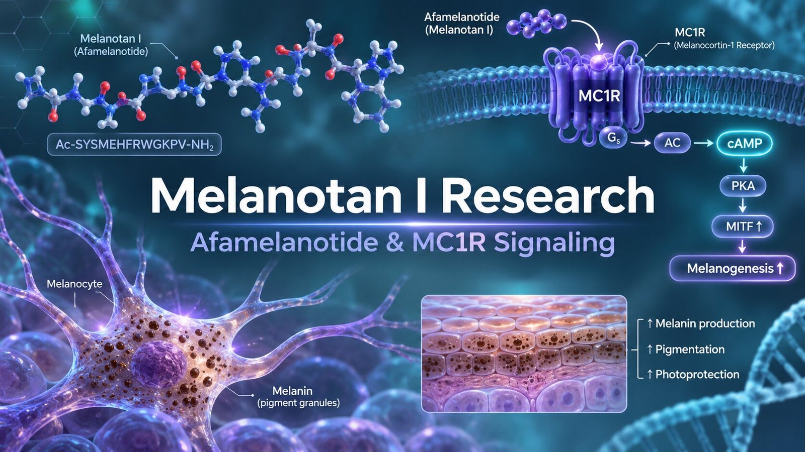
The Basic MC1R Signaling Pathway
A simplified pathway is:
Melanotan I → MC1R → Gs → Adenylyl Cyclase → cAMP
Increased cAMP can activate protein kinase A.
Protein kinase A then influences downstream transcriptional regulators.
One important factor is:
MITF
or:
Microphthalmia-associated transcription factor
MITF regulates genes involved in melanocyte biology and melanin synthesis.
MITF Research
MITF is one of the major transcription factors in melanocytes.
It influences expression of proteins involved in pigmentation.
These include:
- Tyrosinase
- TYRP1
- DCT
Activation of MC1R and cAMP signaling can increase MITF-related activity.
This provides one pathway connecting receptor activation with changes in pigment production.
Tyrosinase
Tyrosinase is a key enzyme in melanin synthesis.
It participates in early steps of the pathway that converts tyrosine into melanin precursors.
Research on MC1R agonists often measures:
- Tyrosinase expression
- Tyrosinase activity
- Melanin content
- cAMP
These measurements help determine whether receptor activation produces downstream melanogenic signaling.
Eumelanin and Pheomelanin
Human skin contains different forms of melanin.
Two major categories are:
- Eumelanin
- Pheomelanin
Eumelanin is generally darker brown or black.
Pheomelanin is more yellow-red.
MC1R activity strongly influences the balance between these pigment pathways.
Functional MC1R signaling favors eumelanin production.
Human Eumelanin Research
An early human study examined [Nle4-D-Phe7]-α-MSH and measured skin pigment composition using analytical methods.
The researchers reported increased eumelanin after experimental exposure while pheomelanin did not show the same significant change.
This provided biochemical evidence that Melanotan I affects the eumelanin pathway.
However, this type of controlled research should not be interpreted as guidance for personal use.
Why D-Phenylalanine Matters
One of the defining structural modifications in Melanotan I is:
D-Phe7
Natural α-MSH contains L-phenylalanine at this position.
Changing the stereochemistry to D-phenylalanine influences:
- Peptide shape
- Enzyme recognition
- Receptor interaction
- Biological stability
Structural research on MC1R has shown that afamelanotide interacts strongly with the receptor and that the D-Phe7 modification contributes to important receptor contacts.
Why Norleucine Matters
Natural α-MSH contains methionine at position 4.
Melanotan I contains:
Norleucine
instead.
Methionine can undergo oxidation.
Replacing it with norleucine helped create a more chemically stable analogue.
This was one of the key steps in the development of NDP-MSH.
Structure-Activity Research
Melanotan I is an important example of peptide structure-activity optimization.
Researchers modified only a small part of the natural peptide while preserving the overall hormone framework.
The goal was to improve:
- Stability
- Potency
- Receptor interaction
This demonstrates how a small number of amino-acid substitutions can substantially alter peptide pharmacology.
The HFRW Motif
Melanocortin peptides contain an important conserved sequence:
His-Phe-Arg-Trp
often abbreviated as:
HFRW
This motif plays a major role in melanocortin receptor recognition.
Structural studies show that the HFRW region occupies the receptor-binding pocket and contributes strongly to MC1R activation.
In Melanotan I, the corresponding phenylalanine is present as D-Phe, altering the geometry of this important pharmacophore.
Modern MC1R Structural Research
Cryo-electron microscopy has provided detailed structures of MC1R bound to several ligands.
These include:
- Natural α-MSH
- Afamelanotide
- SHU9119
The structures revealed:
- The MC1R binding pocket
- Calcium-dependent ligand recognition
- Gs coupling
- Important peptide-receptor contacts
This provides direct structural evidence explaining how afamelanotide activates MC1R.
Calcium Plays a Role in Ligand Recognition
MC1R binding involves an interesting feature:
calcium
Structural research showed that calcium contributes to receptor recognition of melanocortin ligands.
This makes melanocortin receptor pharmacology more complex than a simple peptide-receptor interaction.
Researchers studying receptor binding should therefore consider:
- Ligand sequence
- Receptor structure
- Calcium concentration
- G-protein coupling
Gs Protein Coupling
MC1R is primarily associated with Gs signaling.
When activated:
- Ligand binds MC1R.
- MC1R activates Gs.
- Gs stimulates adenylyl cyclase.
- cAMP increases.
- Protein kinase A signaling increases.
- Melanocyte transcription pathways change.
This pathway provides multiple experimental endpoints.
cAMP Assays
cAMP assays are commonly used to evaluate melanocortin receptor agonists.
Researchers can expose MC1R-expressing cells to different peptide concentrations.
They may then calculate:
- EC50
- Maximum response
- Concentration-response curve
Comparing α-MSH with Melanotan I can reveal changes in receptor potency.
Receptor-Binding Experiments
Researchers may also perform direct binding studies.
Possible methods include:
- Radioligand competition
- Fluorescent ligand assays
- Structural modeling
- Cryo-EM
- Receptor mutagenesis
These approaches can identify which receptor residues are most important for Melanotan I activity.
MC1R Variants
MC1R is genetically variable.
Some human MC1R variants have reduced receptor function.
Certain variants are associated with pigmentation traits, including red hair and lighter skin phenotypes.
This creates an important research variable.
Two cells with different MC1R variants may respond differently to the same melanocortin ligand.
Why Genetic Variation Matters
A peptide may show strong activity in a laboratory cell line expressing one receptor form.
That does not mean the response will be identical across all receptor variants.
Researchers may therefore compare:
- Wild-type MC1R
- Reduced-function MC1R variants
- Receptor knockout controls
This can help define receptor dependence.
Melanocyte Cell Models
Human melanocyte cultures are especially useful for Melanotan I research.
Possible endpoints include:
- cAMP
- MITF expression
- Tyrosinase
- Melanin content
- Oxidative-stress responses
- Receptor expression
These experiments can connect receptor activation with cellular output.
Keratinocyte-Melanocyte Communication
Skin pigmentation is not controlled by melanocytes alone.
Keratinocytes also participate.
They can produce melanocortin-related signals and interact with melanocytes.
Modern MC1R research emphasizes this local skin signaling system.
Researchers studying Melanotan I may therefore use:
- Melanocyte monoculture
- Keratinocyte culture
- Co-culture systems
Each model answers different questions.
Melanosome Research
Melanocytes produce melanin inside specialized organelles called:
melanosomes
These structures are then transferred to keratinocytes.
Researchers can examine:
- Melanosome formation
- Melanin synthesis
- Melanosome transport
- Transfer to keratinocytes
MC1R activation can influence several parts of this system.
Oxidative-Stress Research
MC1R signaling is also associated with cellular responses to oxidative stress.
Recent reviews describe roles for activated MC1R in melanocyte homeostasis and responses to environmental stressors.
Researchers may therefore investigate:
- Reactive oxygen species
- Antioxidant pathways
- DNA damage responses
- Cell survival
This broadens Melanotan I research beyond pigmentation alone.
DNA Damage Response
Ultraviolet radiation can damage DNA.
Human melanocytes respond through several repair and stress-response pathways.
MC1R activation has been linked with improved DNA-damage response in experimental melanocyte systems.
This does not mean a research peptide should be described as a substitute for photoprotection.
The finding is a mechanistic laboratory observation.
Melanotan I Is a Linear Peptide
Melanotan I retains the linear 13-residue α-MSH framework.
This is an important difference from Melanotan II.
Melanotan II is a cyclic peptide analogue.
Cyclization changes:
- Molecular conformation
- Receptor profile
- Stability
- Pharmacology
Melanotan I should therefore not be treated as interchangeable with Melanotan II.
Melanotan I vs Melanotan II
Melanotan I
- Linear 13-residue α-MSH analogue
- [Nle4, D-Phe7]-α-MSH
- Strong research focus on MC1R
- Closely related to afamelanotide
Melanotan II
- Cyclic melanocortin analogue
- Shorter peptide
- Broader melanocortin receptor activity
- Different pharmacological profile
These structural differences create distinct research topics.
Melanotan I vs PT-141
PT-141, also called bremelanotide, is another cyclic melanocortin-related peptide.
Its research focuses more strongly on:
- MC3R
- MC4R
- Central nervous system signaling
Melanotan I instead has a much stronger historical focus on:
- MC1R
- Melanocytes
- Eumelanin
- Pigmentation biology
This keeps the two subjects scientifically separate.
Melanotan I vs KPV
KPV is the tripeptide:
Lys-Pro-Val
It corresponds to the final three residues of α-MSH.
KPV research focuses primarily on:
- Epithelial biology
- Inflammatory pathways
- NF-κB
- PepT1 transport
Melanotan I preserves almost the entire α-MSH framework and strongly engages melanocortin receptor signaling.
They are very different research peptides.
Melanotan I vs Natural Alpha-MSH
Natural α-MSH and Melanotan I share the same basic 13-residue architecture.
The important differences are:
Natural α-MSH
- Met at position 4
- L-Phe at position 7
Melanotan I
- Nle at position 4
- D-Phe at position 7
These changes increase stability and receptor potency.
Pharmacokinetic Research
Older human pharmacokinetic research examined Melanotan I using different administration routes.
One study reported a terminal plasma half-life of approximately 0.8 to 1.7 hours after subcutaneous administration under the experimental conditions.
This historical finding is useful scientifically.
However, it should not be converted into dosing guidance.
Why Pharmacokinetic Data Need Context
Pharmacokinetic measurements depend on:
- Formulation
- Assay method
- Route
- Population
- Sampling schedule
Older studies may also use analytical methods different from modern peptide assays.
Researchers should therefore interpret historical values cautiously.
Afamelanotide Stability Research
A 2026 analytical study specifically examined the stability profile of afamelanotide using liquid chromatography and high-resolution mass spectrometry.
The researchers highlighted the importance of the Nle4 and D-Phe7 substitutions and evaluated degradation behavior using modern analytical methods.
This provides a useful modern reference for laboratory characterization.
Why Stability Matters
Peptides can degrade through several processes.
These may include:
- Oxidation
- Hydrolysis
- Deamidation
- Peptide-bond cleavage
Melanotan I was specifically designed partly to improve resistance to instability associated with native α-MSH.
However, modified peptides still require careful storage and analysis.
Analytical Characterization
Research-grade Melanotan I should be evaluated using appropriate analytical methods.
Useful techniques include:
- HPLC
- LC-MS
- High-resolution mass spectrometry
- Peptide mapping
Researchers should verify both:
- Purity
- Identity
HPLC Analysis
High-performance liquid chromatography can help evaluate:
- Main peptide peak
- Relative purity
- Secondary peaks
- Batch consistency
However, HPLC does not independently establish the exact peptide identity.
Mass Spectrometry
Mass spectrometry is particularly useful for Melanotan I because the molecule includes non-native residue features.
Researchers can use MS to verify:
- Expected molecular mass
- Peptide integrity
- Degradation products
- Synthesis-related impurities
Modern LC-HRMS methods have been used directly to investigate afamelanotide stability.
Confirming the D-Phe Modification
Analytical identity matters because stereochemistry influences activity.
A peptide containing L-Phe rather than D-Phe would not be chemically identical to Melanotan I.
Mass alone may not always distinguish stereoisomers.
Therefore, manufacturing documentation and validated synthesis controls remain important.
Purity Is Not Identity
A sample may produce a chromatogram showing 99% main peak.
That only addresses purity.
Researchers should still confirm:
- Correct peptide
- Correct substitutions
- Correct molecular mass
Purity and identity are separate quality measures.
Certificate of Analysis Review
A useful Melanotan I Certificate of Analysis may include:
- Product name
- Peptide identity
- Lot number
- HPLC purity
- Mass-spectrometry result
- Test date
- Storage information
Batch-specific documentation is preferable to a generic specification sheet.
Storage Considerations
Peptide stability may be affected by:
- Temperature
- Moisture
- Light
- pH
- Oxidizing conditions
- Repeated freeze-thaw cycles
Researchers should follow the product-specific COA and storage documentation.
The stability of lyophilized material may differ from material in solution.
Experimental Design for MC1R Research
A well-designed Melanotan I receptor study may include:
- Vehicle control
- Natural α-MSH
- Melanotan I
- MC1R antagonist
- MC1R knockout control
- Melanotan II comparator
This allows researchers to separate:
- Receptor-specific effects
- Analogue-specific effects
- General melanocortin activity
Concentration-Response Studies
Testing one concentration provides limited information.
Researchers may evaluate multiple concentrations and calculate:
- EC50
- Maximum response
- Relative potency
This is particularly useful when comparing Melanotan I with native α-MSH.
Time-Course Studies
Different endpoints develop at different speeds.
For example:
- cAMP may change rapidly
- MITF may change later
- Melanin accumulation may require longer observation
Researchers should match the sampling time to the biological endpoint.
Cell Viability Controls
Melanocyte experiments should include viability measurements.
Reduced signaling could otherwise reflect:
- Cellular stress
- Toxicity
- Nutrient depletion
Cell viability helps separate pharmacological effects from nonspecific damage.
FDA-Approved Afamelanotide Is a Specific Pharmaceutical Product
Afamelanotide has an important regulatory history.
The FDA-approved product SCENESSE contains afamelanotide and is indicated to increase pain-free light exposure in adult patients with a history of phototoxic reactions from erythropoietic protoporphyria.
This approval applies to a specific regulated implant.
It should not be generalized to research-grade Melanotan I products.
Research Material vs SCENESSE
A research material and an FDA-approved implant differ in important ways.
SCENESSE is manufactured under pharmaceutical controls and supplied as a defined implant formulation.
Research-grade Melanotan I is intended for laboratory investigation.
Researchers should not imply interchangeability based only on shared peptide identity.
Unregulated Melanotan Products Are a Separate Issue
Scientific reviews have raised concerns about unregulated Melanotan products marketed online for tanning.
Reported concerns include:
- Uncertain preparation
- Unverified purity
- Unknown dosing
- Dermatologic changes
- Lack of regulatory oversight
A review specifically warned that unregulated Melanotan I and II products should not be confused with tested afamelanotide pharmaceutical material.
This reinforces the importance of research-only labeling and analytical verification.
Evidence Hierarchy Matters
Melanotan I has several different evidence categories.
Molecular evidence
Includes:
- MC1R structural studies
- Receptor binding
- G-protein coupling
Cellular evidence
Includes:
- Melanocyte signaling
- cAMP
- Melanin production
Human research
Includes:
- Early pharmacokinetic studies
- Pigmentation studies
- Clinical development of afamelanotide
Regulatory evidence
Includes:
- FDA approval of a specific afamelanotide implant
These evidence types answer different questions.
Avoiding Overstatement
Responsible Melanotan I content should avoid statements such as:
- “Melanotan I safely produces tanning”
- “Melanotan I protects everyone from UV”
- “Research-grade Melanotan I is the same as SCENESSE”
More accurate language focuses on:
- MC1R signaling
- Melanocyte models
- Eumelanin research
- Receptor structure
- Peptide stability
Melanotan I Research and Bayoupep USA
Within the Bayoupep USA research catalog, Melanotan I should be presented strictly as a laboratory research peptide.
Researchers evaluating the material should confirm:
- Correct [Nle4, D-Phe7]-α-MSH identity
- Batch-specific purity
- Analytical verification
- Lot traceability
- Storage requirements
For research purposes only. Not for human or veterinary use.
Common Questions About Melanotan I Research
What is Melanotan I?
Melanotan I is a synthetic α-MSH analogue containing Nle4 and D-Phe7 substitutions.
Is Melanotan I the same as afamelanotide?
The peptide structure commonly called Melanotan I or NDP-MSH corresponds to afamelanotide.
Research-grade material, however, is not automatically equivalent to an approved pharmaceutical formulation.
How many amino acids are in Melanotan I?
Melanotan I retains the 13-residue α-MSH framework.
What is the main receptor studied with Melanotan I?
MC1R is the primary receptor of interest in pigmentation and melanocyte research.
What signaling pathway is involved?
MC1R primarily activates:
Gs → Adenylyl Cyclase → cAMP
which influences downstream melanocyte signaling.
Why is D-Phe7 important?
D-Phe7 improves structural stability and contributes to strong receptor interactions.
Why is norleucine used?
Norleucine replaces methionine at position 4, helping reduce problems associated with methionine oxidation.
Is Melanotan I the same as Melanotan II?
No.
Melanotan I is a linear 13-residue α-MSH analogue.
Melanotan II is a different cyclic melanocortin peptide.
Is Melanotan I the same as PT-141?
No.
PT-141 is another cyclic melanocortin analogue with a different structure and receptor emphasis.
Is afamelanotide FDA approved?
A specific afamelanotide implant, SCENESSE, is FDA approved for a defined erythropoietic protoporphyria indication.
That does not make research-grade Melanotan I an approved pharmaceutical product.
Final Thoughts
Melanotan I is a structurally modified analogue of alpha-melanocyte-stimulating hormone and one of the most important research peptides in melanocortin biology.
Its defining structure is:
[Nle4, D-Phe7]-α-MSH
These substitutions distinguish it from native α-MSH and contribute to increased stability and receptor activity.
The strongest research areas include:
- MC1R receptor activation
- Gs signaling
- cAMP production
- MITF regulation
- Tyrosinase-related pathways
- Eumelanin synthesis
- Melanocyte biology
- Peptide stability
- Structure-activity relationships
Modern cryo-EM research has directly visualized afamelanotide bound to MC1R, revealing the receptor pocket, calcium-dependent recognition, and G-protein activation mechanism.
Melanotan I is also scientifically distinct from Melanotan II and PT-141.
Melanotan I preserves the linear α-MSH structure and is strongly associated with MC1R and melanocyte research. Melanotan II and PT-141 are cyclic peptides with different receptor profiles and research applications.
Researchers should also distinguish research-grade Melanotan I from the FDA-approved afamelanotide implant SCENESSE. The pharmaceutical product has defined manufacturing, formulation, quality, and regulatory controls that should not be assumed for unrelated research materials.
For laboratory work, strong experimental practice should include:
- Exact peptide identity verification
- Confirmation of Nle4 and D-Phe7 modifications
- HPLC purity analysis
- Mass-spectrometry testing
- MC1R-specific controls
- cAMP measurements
- Appropriate melanocyte models
- Batch traceability
Melanotan I is therefore best understood as a synthetic linear α-MSH analogue used to investigate melanocortin receptor biology, MC1R signaling, melanocyte function, and peptide structure-activity relationships.
This information is provided for scientific and educational research purposes only. Melanotan I research materials are not intended for human or veterinary use.