
In this article
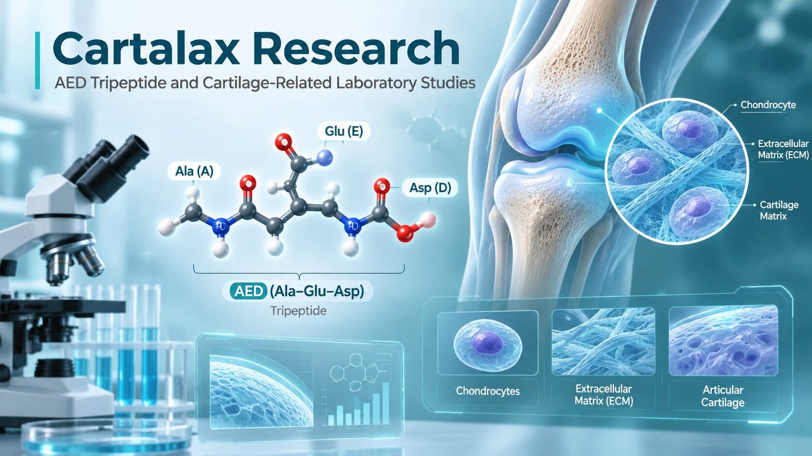
Cartalax is a short synthetic peptide most commonly identified in research literature as the AED peptide or Ala-Glu-Asp.
It contains only three amino-acid residues:
- Alanine
- Glutamic acid
- Aspartic acid
Because it contains three residues, Cartalax is classified as a tripeptide.
Cartalax belongs to a group of short peptides studied within the peptide bioregulator research program associated with the St. Petersburg Institute of Bioregulation and Gerontology. In scientific papers, the peptide may appear under names such as:
- AED
- T-31
- Ala-Glu-Asp
- Alanyl-glutamyl-aspartic acid
Cartalax is often marketed in connection with cartilage and joint research. However, its evidence base requires careful interpretation.
Recent peer-reviewed studies have examined AED in chondrocytes and human mesenchymal stem-cell models, including experiments related to chondrogenic differentiation and cellular aging. These are laboratory studies, not proof that the peptide regenerates cartilage or treats osteoarthritis in humans.
The published Cartalax/AED literature is also relatively small and heavily concentrated within one research network. Independent replication remains limited.
For that reason, Cartalax is best approached as an experimental short peptide for laboratory research, rather than as an established cartilage therapy.
For research purposes only. Not for human or veterinary use.
What Is Cartalax?
Cartalax is generally identified as the synthetic tripeptide:
Ala-Glu-Asp
This sequence is commonly abbreviated:
AED
The peptide has also appeared in the literature under the research code:
T-31
Three-residue peptides are much smaller than many well-known signaling peptides.
For comparison:
- KPV contains three residues
- GHK contains three residues
- Glutathione contains three amino-acid components
- Melanotan I contains 13 residues
- Tesamorelin contains 44 residues
A short sequence does not necessarily mean a simple biological effect.
Even very small peptides can potentially interact with:
- Proteins
- Enzymes
- Cellular membranes
- Regulatory pathways
However, those interactions must be demonstrated experimentally.
Why Cartalax Is Associated With Cartilage Research
Cartalax is commonly described as a cartilage-related peptide because AED has been investigated in experimental models involving:
- Chondrocytes
- Mesenchymal stem cells
- Chondrogenic differentiation
- Cellular aging
- Extracellular-matrix markers
A 2023 study evaluated AED in human mesenchymal stem cells undergoing replicative aging.
Researchers examined several markers involved in chondrogenesis, including:
- SOX9
- Aggrecan
- Type II collagen
- COMP
The study reported increased gene expression and protein synthesis of these markers under the tested cell-culture conditions.
These findings provide a laboratory basis for continued cartilage-related research.
They do not demonstrate clinical cartilage repair.
Chondrocytes Are Central to Cartilage Biology
Chondrocytes are the primary resident cells of cartilage.
They are responsible for maintaining the cartilage extracellular matrix.
This matrix contains important structural components such as:
- Type II collagen
- Aggrecan
- Proteoglycans
- Water-binding glycosaminoglycans
Healthy cartilage depends on a balance between:
- Matrix production
- Matrix degradation
- Cell survival
- Mechanical stress responses
Cartilage degeneration can develop when this balance shifts.
Researchers therefore examine whether experimental peptides influence chondrocyte activity or matrix-related pathways.
What Is Chondrogenesis?
Chondrogenesis is the biological process through which cells develop toward a cartilage-producing phenotype.
Mesenchymal stem cells can be used experimentally to study this process.
During chondrogenic differentiation, researchers often examine markers such as:
- SOX9
- COL2A1
- Aggrecan
- COMP
These markers provide information about whether cells are moving toward a cartilage-related phenotype.
The 2023 AED study reported changes in several of these markers during replicative aging of human mesenchymal stem cells.
SOX9 Research
SOX9 is one of the most important transcription factors in cartilage development.
It helps regulate genes involved in the chondrocyte phenotype.
SOX9 is strongly connected with:
- Type II collagen
- Aggrecan production
- Chondrogenic differentiation
Researchers frequently use SOX9 as an experimental marker when investigating cartilage formation.
An increase in SOX9-related activity can suggest movement toward a chondrogenic state.
However, one molecular marker cannot independently prove new cartilage formation.
Type II Collagen
Type II collagen is one of the main structural proteins in articular cartilage.
It forms an important fibrillar framework within the extracellular matrix.
Researchers often measure:
- COL2A1 gene expression
- Type II collagen protein
when studying chondrocyte differentiation.
The 2023 AED study reported increased type II collagen-related expression and protein synthesis in its mesenchymal stem-cell model.
This is a useful mechanistic observation.
It should not be converted into a claim that Cartalax rebuilds damaged joints.
Aggrecan
Aggrecan is a large proteoglycan found in cartilage.
It helps cartilage retain water and resist compression.
Loss of aggrecan is commonly associated with cartilage degeneration.
Chondrogenic research therefore often includes aggrecan as a laboratory endpoint.
AED-associated changes in aggrecan expression have been reported in experimental stem-cell differentiation models.
COMP
COMP stands for:
Cartilage Oligomeric Matrix Protein
It is an extracellular-matrix protein found in cartilage and related tissues.
Researchers may measure COMP during studies involving:
- Chondrocyte function
- Matrix formation
- Cartilage differentiation
The AED mesenchymal stem-cell study included COMP among its measured chondrogenic markers.
Cartalax and Aging Chondrocytes
Another 2023 study examined AED in chondrocytes and focused on cellular aging.
The researchers investigated the senescence-associated secretory phenotype, often abbreviated:
SASP
The study reported age-associated changes involving:
- p16
- p21
- p53
- TNF-α
- IL-1α
- Sirt1
AED exposure altered several of these markers under the experimental conditions.
This makes cellular aging an important part of Cartalax research.
What Is Cellular Senescence?
Cellular senescence is a state in which a cell stops dividing but remains metabolically active.
Senescent cells can release signaling molecules that influence nearby cells.
This secretory pattern is commonly called:
SASP
SASP can include:
- Cytokines
- Chemokines
- Proteases
- Growth-related molecules
In cartilage research, senescent chondrocytes are of interest because their signaling may contribute to age-related changes in the extracellular environment.
p16 and p21
p16 and p21 are proteins commonly associated with cell-cycle control and senescence.
Their expression can increase when cells enter a senescent state.
Researchers may therefore measure these proteins when studying:
- Replicative aging
- Cellular stress
- Chondrocyte senescence
Changes in p16 or p21 do not automatically indicate improved tissue health.
They are markers that must be interpreted within a broader experimental framework.
p53 Research
p53 plays a central role in:
- DNA-damage responses
- Cell-cycle arrest
- Apoptosis
- Stress signaling
AED has also been studied in other tissue models where p53-related changes were observed.
For example, research involving kidney tissue cultures found that T-31, identified as AED, influenced markers of proliferation and apoptosis.
These findings show that AED research is not limited to cartilage.
Sirt1
Sirt1 is a NAD+-dependent deacetylase.
It is widely studied in:
- Cellular aging
- Metabolism
- Stress responses
- Inflammation
The 2023 chondrocyte study reported age-related changes in Sirt1 and examined whether AED affected this pattern.
This gives researchers another possible mechanistic endpoint.
However, changes in Sirt1 alone do not establish a clinical anti-aging effect.
Inflammatory Cytokines in Cartilage Models
Inflammatory signaling is an important component of cartilage research.
Commonly studied cytokines include:
- IL-1
- TNF-α
- IL-6
These molecules can influence:
- Matrix degradation
- Chondrocyte metabolism
- Protease production
The AED chondrocyte study examined TNF-α and IL-1α as part of its SASP analysis.
This suggests one possible research direction for Cartalax.
Extracellular Matrix Biology
Cartilage function depends heavily on its extracellular matrix.
The matrix contains:
- Collagen fibers
- Proteoglycans
- Glycosaminoglycans
- Water
- Matrix-associated proteins
Chondrocytes continuously regulate this environment.
Researchers studying Cartalax may therefore measure both:
Anabolic markers
- Type II collagen
- Aggrecan
- COMP
and
Catabolic markers
- Matrix metalloproteinases
- Inflammatory cytokines
- Senescence-related proteins
This provides a more complete picture.
Matrix Metalloproteinases
Matrix metalloproteinases are commonly abbreviated:
MMPs
These enzymes can break down extracellular-matrix proteins.
Several MMPs are relevant to connective-tissue research.
AED-related research in other tissue systems has examined matrix-remodeling proteins, including MMP-related pathways. A kidney cell-culture study involving T-31 evaluated cellular renewal and extracellular-matrix signaling markers.
Direct Cartalax-specific cartilage evidence involving MMP regulation remains much more limited.
Why Cartilage Is Difficult to Repair
Articular cartilage has a limited natural capacity for repair.
Several biological factors contribute to this problem.
Cartilage:
- Contains relatively few cells
- Has no direct blood supply
- Has limited cell migration
- Experiences continuous mechanical stress
Modern cartilage-regeneration research therefore investigates many approaches, including peptide-based biomaterials and signaling molecules.
Cartalax represents one small area within this broader research field.
Cartalax Is Not the Same as a Cartilage Scaffold Peptide
An important distinction should be made between Cartalax and engineered peptide biomaterials.
Some research peptides are designed to:
- Self-assemble
- Form hydrogels
- Bind collagen
- Act as tissue scaffolds
Cartalax is not primarily described as a structural scaffold.
It is investigated as a small regulatory peptide.
This makes its proposed research role different from self-assembling cartilage peptides.
Mesenchymal Stem Cells
Mesenchymal stem cells, or MSCs, are frequently used in cartilage research because they can differentiate toward multiple connective-tissue lineages.
These may include:
- Bone
- Cartilage
- Fat
Researchers can expose MSCs to experimental compounds and measure changes in chondrogenic markers.
This was the strategy used in the 2023 AED chondrogenic differentiation study.
Replicative Aging
Cells cultured repeatedly can gradually develop aging-related characteristics.
This process is called:
Replicative aging
It provides a laboratory model for studying changes that occur as cells undergo many divisions.
The 2023 AED study specifically examined human mesenchymal stem cells during replicative aging.
This is different from studying cartilage damage inside a living joint.
Cartalax Research Outside Cartilage
One reason Cartalax requires careful interpretation is that AED has also been studied in tissues unrelated to cartilage.
Published experiments include models involving:
- Kidney cells
- Skin fibroblasts
- Mesenchymal stem cells
In kidney tissue cultures, AED influenced markers related to cell renewal and apoptosis.
In human skin fibroblasts, AED was associated with changes in sirtuin expression and collagen I under replicative aging conditions.
These findings suggest that the peptide’s proposed biology may not be strictly cartilage-specific.
Skin Fibroblast Research
Fibroblasts produce extracellular-matrix proteins such as collagen.
A study comparing KE and AED peptides in aging human skin fibroblasts reported that AED increased:
- Sirtuin-1
- Sirtuin-6
- Collagen I
under the tested conditions.
This is scientifically interesting because it shows AED activity in another connective-tissue cell type.
However, collagen I in skin is not equivalent to collagen II in cartilage.
The findings should remain separate.
Kidney Research
Several AED publications involve kidney-related models.
One study reported that T-31 increased Ki-67 and reduced p53-related signals in kidney tissue cultures from young and old animals.
Another investigated signaling markers during in-vitro aging of renal cell cultures.
An animal study also examined AED and related peptides in a model of cisplatin-induced kidney injury.
These findings broaden the research background of AED but do not establish cartilage-specific efficacy.
Tissue-Specific Claims Require Caution
Cartalax is often marketed as though it acts specifically on cartilage.
The peer-reviewed literature does not yet establish a highly selective molecular mechanism directing AED exclusively toward cartilage.
Studies in:
- Kidney cells
- Skin fibroblasts
- Stem cells
- Chondrocytes
suggest broader cellular effects may be possible.
Researchers should therefore avoid assuming tissue specificity without direct evidence.
The Peptide Bioregulator Hypothesis
Cartalax belongs to a research tradition sometimes called short peptide bioregulation.
This concept proposes that very short peptides may influence cellular regulation, including gene expression.
Some studies from this research group have proposed effects involving:
- Nuclear entry
- DNA interaction
- Transcriptional regulation
However, Cartalax-specific molecular mechanisms remain incompletely established.
These ideas should therefore be treated as research hypotheses rather than settled biological facts.
Gene-Expression Research
Gene-expression analysis is especially useful for Cartalax because several reported effects involve changes in proteins and transcriptional markers.
Researchers may measure:
- SOX9
- COL2A1
- ACAN
- COMP
- SIRT1
- p16
- p21
- p53
Methods can include:
- RT-qPCR
- RNA sequencing
- Immunocytochemistry
- Western blotting
Using both RNA and protein measurements provides stronger evidence than either alone.
Gene Expression Does Not Equal Tissue Regeneration
This distinction is critical.
An increase in cartilage-related gene expression does not automatically mean:
- New cartilage forms in a joint
- Mechanical strength improves
- Osteoarthritis reverses
Gene expression is an early mechanistic endpoint.
Additional research would be needed to demonstrate:
- Matrix formation
- Tissue architecture
- Mechanical performance
- In-vivo durability
Protein-Level Validation
If AED increases a cartilage-related gene, researchers should also determine whether the corresponding protein changes.
For example:
COL2A1 gene → Type II collagen protein
or:
ACAN gene → Aggrecan protein
The 2023 MSC study reported both gene-expression and protein-related changes, which strengthens the cell-culture observation.
However, this is still an in-vitro model.
Cartilage Matrix Measurements
Future Cartalax experiments could measure:
- Type II collagen
- Aggrecan
- Glycosaminoglycan content
- COMP
- Matrix deposition
These endpoints would help determine whether molecular changes translate into extracellular-matrix production.
Mechanical Testing Would Be a Stronger Endpoint
Cartilage is a mechanical tissue.
A regenerated matrix must withstand:
- Compression
- Shear
- Repeated loading
Therefore, advanced tissue-engineering studies may examine:
- Compression modulus
- Matrix stiffness
- Structural integrity
Cartalax research has not yet established this type of evidence.
Cartalax and Osteoarthritis Research
Osteoarthritis is a complex joint disorder involving more than cartilage alone.
It can affect:
- Articular cartilage
- Subchondral bone
- Synovium
- Ligaments
- Joint inflammation
A peptide that changes one chondrocyte marker does not automatically address the entire disease process.
Cartalax research should therefore focus on defined cellular mechanisms rather than broad osteoarthritis treatment claims.
Current Evidence for Osteoarthritis Is Limited
Peer-reviewed Cartalax/AED studies include laboratory research related to chondrogenesis and aging chondrocytes.
A review from the originating research network also discusses broader osteoarthritis-related claims.
However, independently replicated, well-controlled human clinical trials of Cartalax have not been established in the indexed evidence reviewed here.
This is a major limitation.
Evidence Concentration Matters
Much of the AED literature comes from researchers associated with the same institutional network.
Independent replication is important because it tests whether:
- Results can be reproduced elsewhere
- Methods produce similar outcomes
- Effects remain consistent across models
Until broader replication occurs, conclusions should remain cautious.
Cartalax and Cellular Proliferation
AED has been associated with changes in proliferation markers in some laboratory systems.
Ki-67 is one commonly used proliferation marker.
Research involving kidney tissue cultures reported increased Ki-67 alongside changes in p53 after AED exposure.
Researchers could examine similar endpoints in cartilage-related cells.
However, increased proliferation is not always beneficial.
The biological context matters.
Cartalax and Apoptosis
Apoptosis is programmed cell death.
Markers used in apoptosis research can include:
- p53
- Caspases
- BAX
- BCL-2
Cartalax-related studies have examined p53 and other cell-survival signals in aging models.
Future cartilage-specific experiments could examine whether AED influences chondrocyte survival under controlled stress conditions.
Cartalax and Senescence
Senescence may be particularly relevant to cartilage aging.
Potential endpoints include:
- p16
- p21
- SASP cytokines
- Sirtuins
The 2023 chondrocyte study provides preliminary evidence that AED can alter some of these markers in vitro.
This makes cellular senescence one of the stronger mechanistic directions for future Cartalax research.
Experimental Design for Cartalax Research
A useful cell-culture study could include:
- Untreated control
- Vehicle control
- AED peptide
- Positive chondrogenic control
- Aging or stress condition
Researchers may then evaluate:
- Cell viability
- SOX9
- Type II collagen
- Aggrecan
- COMP
- p16
- p21
- Cytokines
This would provide both chondrogenic and aging-related data.
Dose-Response Experiments
Researchers should generally use multiple experimental concentrations.
This can help determine:
- Minimum active concentration
- Maximum response
- Toxicity
- Non-linear effects
A single concentration gives only limited information.
Time-Course Studies
Different cellular effects occur at different times.
For example:
- Signaling changes may appear quickly
- Gene expression may change later
- Protein accumulation requires more time
- Matrix deposition may take days
Researchers should therefore match sampling time to the endpoint.
Cell Viability Is Essential
Any apparent biological improvement must be interpreted alongside cell viability.
Useful tests may include:
- ATP-based assays
- Resazurin assays
- Membrane-integrity tests
A change in gene expression is difficult to interpret if the peptide also changes cell survival.
Primary Chondrocytes vs Cell Lines
Primary chondrocytes may represent cartilage biology more closely than immortalized cell lines.
However, primary cells can be:
- More variable
- More difficult to culture
- Donor dependent
Cell lines offer consistency but may behave differently from native tissue.
Strong research may use more than one model.
Young vs Aged Cell Models
Because much Cartalax research involves cellular aging, comparing young and aged cells may be valuable.
Researchers could test whether AED produces different responses in:
- Early-passage cells
- Replicatively aged cells
- Senescence-induced models
This could help determine whether effects are age-state dependent.
Three-Dimensional Cartilage Models
Two-dimensional cell culture is useful but limited.
Three-dimensional systems can better model:
- Cell-matrix interactions
- Tissue architecture
- Diffusion
- Mechanical behavior
Future Cartalax research could use:
- Cartilage organoids
- Hydrogel cultures
- Scaffold-based systems
This would move the evidence closer to tissue-level biology.
Animal Models
Animal osteoarthritis models can provide information about:
- Tissue structure
- Inflammation
- Mechanical function
- Cartilage thickness
However, animal results still do not prove human efficacy.
Peer-reviewed Cartalax-specific joint studies remain limited compared with the much broader cartilage research literature.
Human Research Gap
The biggest missing evidence category is rigorous human research.
For Cartalax, strong human evidence would require studies with:
- Defined product identity
- Appropriate controls
- Predefined outcomes
- Adequate sample size
- Transparent safety reporting
Without such data, clinical claims should remain avoided.
Cartalax vs KPV
Cartalax and KPV are both tripeptides.
Their sequences are completely different.
Cartalax
Ala-Glu-Asp
Main research interests:
- Chondrogenesis
- Cellular aging
- Gene expression
KPV
Lys-Pro-Val
Main research interests:
- Epithelial biology
- NF-κB signaling
- Inflammatory pathways
They should not be grouped simply because both are short peptides.
Cartalax vs GHK-Cu
GHK-Cu is another small peptide-related molecule.
Its main research areas include:
- Copper binding
- Fibroblast biology
- Extracellular matrix
- Gene expression
Cartalax does not contain a copper-binding histidine motif like GHK.
Its research history and proposed mechanism are different.
Cartalax vs BPC-157
BPC-157 is a much longer peptide than AED.
Its research literature often focuses on:
- Experimental injury models
- Gastrointestinal systems
- Tissue-repair pathways
Cartalax is a tripeptide primarily discussed in relation to:
- Cellular regulation
- Chondrocyte aging
- Chondrogenic differentiation
These compounds should not be presented as interchangeable cartilage-repair agents.
Cartalax vs Glutathione
Both Cartalax and glutathione contain three amino-acid components.
However, glutathione is a central endogenous redox molecule.
Its main biology involves:
- GSH/GSSG balance
- Peroxide metabolism
- Thiol chemistry
- Detoxification
Cartalax is instead an experimental regulatory tripeptide.
Their functions are fundamentally different.
Analytical Characterization of Cartalax
Research-grade Cartalax should be analytically characterized.
Useful methods may include:
- HPLC
- LC-MS
- Mass spectrometry
- Peptide identity testing
Because AED is very small, analytical methods should clearly distinguish it from:
- Starting amino acids
- Truncated synthesis products
- Other short peptides
HPLC Analysis
High-performance liquid chromatography can help evaluate:
- Main peptide peak
- Relative purity
- Secondary impurities
- Batch consistency
However, an HPLC purity percentage does not independently confirm the sequence.
Mass Spectrometry
Mass spectrometry can help verify:
- Expected molecular mass
- Peptide integrity
- Synthesis-related impurities
For a tripeptide, even a one-residue error produces a different molecule.
Identity testing is therefore essential.
Sequence Verification
Cartalax should be identified as:
Ala-Glu-Asp
or:
AED
Researchers should avoid confusing it with similar short peptides.
In particular, it should not be confused with:
AEDG
which is Epithalon.
Adding one glycine residue changes the molecular identity completely.
Cartalax vs Epithalon
This distinction is especially important.
Cartalax
AED
Ala-Glu-Asp
Three residues.
Epithalon
AEDG
Ala-Glu-Asp-Gly
Four residues.
They belong to related short-peptide research traditions but are different molecules with different research contexts.
Purity Is Not the Same as Identity
A sample may show a high purity percentage yet still contain the wrong peptide.
Researchers should ask two questions:
Purity:
How much of the detectable material is the main component?
Identity:
Is the main component actually AED?
Both should be verified.
Certificate of Analysis Review
A useful Cartalax Certificate of Analysis may include:
- Product identity
- AED sequence
- Batch number
- HPLC purity
- Mass-spectrometry result
- Testing date
- Storage recommendations
Batch-specific documentation is more useful than a generic specification.
Storage Considerations
Small peptides may be affected by:
- Moisture
- Temperature
- pH
- Contamination
- Repeated environmental changes
Researchers should follow the storage instructions provided for the specific research material.
Dry peptide and prepared solutions may have different stability profiles.
Research Material vs Commercial Claims
Cartalax is sold or discussed online with strong claims involving:
- Cartilage repair
- Joint regeneration
- Anti-aging effects
The published evidence does not support presenting these statements as established outcomes.
The strongest current data remain:
- Cell-culture studies
- Aging-related markers
- Chondrogenic differentiation markers
- Limited preclinical work
Responsible scientific content should reflect that distinction.
What the Evidence Currently Supports
A careful summary is:
AED has shown measurable effects on gene expression, protein synthesis, cellular aging markers, and chondrogenic markers in selected experimental models.
That is supportable from the available studies.
The evidence does not establish that Cartalax:
- Rebuilds human cartilage
- Reverses osteoarthritis
- Prevents joint degeneration
- Produces proven clinical benefits
Those claims would go beyond the current evidence.
Useful Future Research Questions
Cartalax research could be strengthened by answering several questions:
- Does AED directly influence human articular chondrocytes?
- Are results reproducible across independent laboratories?
- Does AED increase functional cartilage matrix production?
- Does it change matrix-degrading enzymes?
- Does it influence mechanical tissue properties?
- What molecular target binds AED?
- Is there a receptor?
- Does AED enter the nucleus?
- Are effects sequence-specific?
- Do animal cartilage findings translate across species?
These questions remain open.
Common Questions About Cartalax
What is Cartalax?
Cartalax is commonly identified as the synthetic tripeptide Ala-Glu-Asp, abbreviated AED.
How many amino acids does Cartalax contain?
Three.
It is therefore a tripeptide.
Is Cartalax the same as Epithalon?
No.
Cartalax is AED.
Epithalon is AEDG.
Has Cartalax been studied in cartilage-related cells?
Yes.
Recent studies have examined AED in aging chondrocytes and chondrogenic differentiation of human mesenchymal stem cells.
What cartilage markers have been studied?
Reported experimental markers include:
- SOX9
- Aggrecan
- Type II collagen
- COMP
Has Cartalax been proven to regenerate human cartilage?
No.
Current peer-reviewed evidence does not establish clinical cartilage regeneration.
Is there strong independent replication?
Independent replication remains limited.
Much of the AED research comes from the same research network.
Is Cartalax FDA approved?
Cartalax should not be presented as an FDA-approved therapeutic drug.
What should researchers verify in a Cartalax sample?
Researchers should confirm:
- AED identity
- Correct sequence
- Purity
- Molecular mass
- Batch traceability
- Storage conditions
Final Thoughts
Cartalax is a very short peptide, but its research story is more complex than its three-amino-acid structure might suggest.
The peptide is generally identified as:
Ala-Glu-Asp, or AED.
Its most relevant modern laboratory research involves two areas:
- Chondrogenic differentiation
- Cellular aging in chondrocytes
A 2023 human mesenchymal stem-cell study reported AED-associated increases in markers including SOX9, aggrecan, type II collagen, and COMP during replicative aging.
Another study examined aging-associated signaling in chondrocytes and reported changes involving p16, p21, p53, inflammatory cytokines, and Sirt1 following AED exposure.
AED has also been studied in kidney and skin cell models, showing that its experimental history extends beyond cartilage alone.
These findings provide a basis for further research.
They do not establish Cartalax as a proven cartilage-regeneration treatment.
The strongest future studies would combine:
- Human primary chondrocytes
- Independent replication
- Dose-response testing
- Matrix measurements
- Senescence markers
- Three-dimensional cartilage models
- Mechanical tissue testing
- HPLC and mass-spectrometry verification
Cartalax is therefore best understood as an experimental AED tripeptide being investigated for effects on cellular regulation, chondrogenic differentiation, extracellular-matrix markers, and aging-related pathways in laboratory models.
This information is provided for scientific and educational research purposes only. Cartalax research materials are not intended for human or veterinary use.